
News | June 2, 2026
Keratosis often seems harmless at first: a rough patch on the forehead, nose, scalp, back of the hand, or forearm that feels like fine sandpaper. It is precisely this sensation that leads many people to seek a dermatologist’s consultation. This is a wise move, because while keratosis does not always indicate skin cancer, the actinic form is a serious form of UV-induced skin damage.
At the ROC Derma private practice in Aschheim near Munich, we medically evaluate such skin changes, assess risk factors, and discuss which treatment is best suited to your findings, skin type, daily routine, and previous treatments. This guide explains how to recognize keratosis, the different types that exist, and when treatment for actinic keratosis is recommended.
The term "keratosis" refers to an increase in the keratinization of the skin. Keratin, a structural protein in the epidermis, protects the skin in everyday life. If the skin's natural renewal process becomes disrupted, rough, scaly, thickened, or crusty areas may develop. The causes range from the aging process and friction to long-term exposure to UV radiation.
For patients, one question matters above all else: Is the lesion benign, does it require monitoring, or does it require treatment? No search engine or photo comparison can provide the answer. A doctor evaluates the color, surface, borders, texture, location, pattern, and personal risk factors. If necessary, a reflected-light microscope is used to supplement the examination.
Keratosis in sun-exposed areas warrants special attention. UV damage accumulates over the years. People who play a lot of golf, sail, ski, garden, jog, or work outdoors for a living often have a high lifetime dose of UV exposure in their skin.
Actinic keratosis is caused by chronic exposure to UV radiation. It primarily affects the so-called “sun-exposed areas”: the forehead, nose, cheeks, ears, hairless scalp, lips, décolleté, forearms, and the backs of the hands. Many people do not initially experience any pain, but rather notice a patch of skin that remains rough despite skincare.
Typical symptoms include skin-colored, reddish, or brownish patches with fine scaling. Later, the skin feels firmer, rougher, or more crusty. On the scalp, keratosis becomes noticeable when washing, shaving, or combing. On the lower lip, UV damage often appears as a dry, cracked, flaky area.
In dermatology, actinic keratosis is considered an early form or precursor of squamous cell carcinoma of the skin. This does not mean that every single lesion will necessarily develop into skin cancer. It means that the skin shows cellular changes that require medical evaluation and, in most cases, treatment.
Anyone searching for “keratosis images” or “actinic keratosis images” will find a wide variety of skin conditions. Comparing them is only helpful to a limited extent. Many changes look similar but behave differently. This overview shows the most common classifications:
| Form | Typical characteristics | Medical Classification |
|---|---|---|
| Actinic keratosis | Rough, scaly, reddish, or skin-colored patches on sun-exposed skin | UV-induced cellular changes associated with an increased risk of non-melanoma skin cancer |
| Seborrheic keratosis | A brownish, somewhat warty surface that appears "stuck on" | Usually a benign age-related change; consult a dermatologist if in doubt |
| Friction-induced calluses | Thickened skin on pressure points, hands, or feet | Mechanical cause; treatment depends on the trigger |
| Keratosis pilaris | Small, rough nodules, often on the upper arms or thighs | Benign keratinization disorder of the hair follicles |
Accurate classification is important because the treatment of keratosis depends on the type. A cosmetically bothersome seborrheic keratosis requires a different approach than actinic keratosis on the scalp with multiple areas of UV damage in the surrounding area.
Make an appointment with a dermatologist if a rough patch persists for several weeks, grows, bleeds, oozes, hurts, or returns after treatment. The same applies to scabs that fall off and then reappear. Keratosis with a palpable thickening, inflammation, or unclear borders requires medical evaluation.
Special caution is required with regard to these risk factors:
Given these circumstances, a cursory examination is not enough. At the ROC, we combine a dermatological examination with a structured risk assessment. If other medical specialties appear relevant—for example, in cases of comorbidities or chronic inflammatory conditions—patients benefit from the Regenerative Center’s interdisciplinary approach.
Dermatologists can often clinically identify a typical actinic keratosis. The doctor examines the entire affected area, palpates the surface, and checks for signs of field cancerization in the surrounding area. This term describes a sun-damaged area of skin with multiple visible or palpable changes.
Dermatoscopy is used when findings are unclear. It reveals structures that cannot be reliably assessed with the naked eye. If a lesion does not fit the diagnosis, is heavily keratinized, remains resistant to treatment, or shows signs of squamous cell carcinoma, a tissue sample can provide clarity.
Search queries such as “early-stage actinic keratosis images” or “grade 3 actinic keratosis images” reveal a clear need: those affected want to assess their risk. Images are no substitute for a medical examination. Factors such as light, skin type, inflammation, skincare products, and scratching can alter the appearance of the condition. The progression of the condition often provides more information than a single snapshot.
Treatment for actinic keratosis depends on the number, thickness, location, and extent of the lesions. Dermatologists often treat individual small lesions on a lesion-by-lesion basis. If there are multiple lesions or an area of UV-damaged skin, a field-based therapy is often appropriate.
Common treatments include cryotherapy, surgical excision, laser therapy, photodynamic therapy, and topical medications. An actinic keratosis ointment may contain active ingredients such as 5-fluorouracil, imiquimod, diclofenac, or tirbanibulin. The choice of substance depends on the clinical findings, regulatory approval, tolerability, duration of treatment, and inflammatory response.
What kind of skin reaction can you expect? Redness, a burning sensation, flaking, and scabbing may be part of the intended effect during treatment with the ointment. Before you begin, we’ll explain what is normal, when a follow-up visit is recommended, and how to care for your skin during the healing phase.
Cryotherapy is often used for clearly localized lesions. Liquid nitrogen specifically damages the abnormal cells; the area forms a scab and usually heals within a few days to a few weeks. More severely thickened areas can be removed or, in cases of diagnostic uncertainty, examined histologically.
Laser treatments can remove superficial hyperkeratosis and, depending on the findings, are suitable for treating individual lesions or larger areas. For more information, please visit the ROC Derma website.
If there are multiple rough patches on the forehead, scalp, or back of the hands, it is often not just a single case of keratosis. The surrounding tissue has likely sustained UV damage that is not yet fully visible. A targeted treatment addresses precisely this area.
Photodynamic therapy combines a light-activated agent with a specific light exposure. Damaged cells react more strongly than healthy skin cells. For suitable conditions, this procedure offers the advantage of detecting visible and early changes within a treatment area.
Photodynamic therapy (PDT) is a proven treatment method for actinic keratoses and is particularly suitable for larger areas of skin with multiple sun-induced cellular changes. The procedure begins with the application of a special active ingredient to the affected skin areas, which accumulates preferentially in the altered cells.
In conventional PDT, the active ingredient is activated using a special medical light source after a certain exposure time. This allows for targeted treatment of the damaged cells while largely sparing the surrounding healthy tissue.
Another form of treatment is daylight PDT (Daylight-PDT). In this method, the active ingredient is activated not by a special light source, but by natural daylight. After the active ingredient is applied, patients spend a set amount of time outdoors. This method is particularly suitable for certain forms of actinic keratosis and can be performed when weather conditions are favorable.
At ROC Derma, we combine PDT with modern laser procedures as needed. Targeted pre-treatment of the skin can enhance the absorption of the active ingredient and further optimize the treatment of sun-induced skin damage. At the same time, many patients benefit from a visible improvement in their skin’s appearance, as other light-induced skin changes—in addition to actinic keratoses—can also be positively influenced. This often allows for the effective combination of medical treatment and aesthetic skin rejuvenation.
Scalp keratosis often affects people with thinning hair or bald spots. Over the course of decades, this area is exposed to UV radiation, often without adequate protection. Wearing a cap while exercising isn’t enough if it’s rarely worn; many people forget to apply sunscreen to their scalp.
These typically appear as several small, rough patches. Shaving can cause minor cuts that bleed or form scabs. This is exactly when it’s worth making an early appointment. A thin actinic keratosis is usually easier to treat than thick, recurring scaly patches.
If you are coming from Munich, Aschheim, or the surrounding area, please plan your visit using the directions to the ROC in Aschheim. Please bring any previous medical reports, a list of medications, and photos documenting the progression of the condition if the affected skin area has changed in recent weeks.
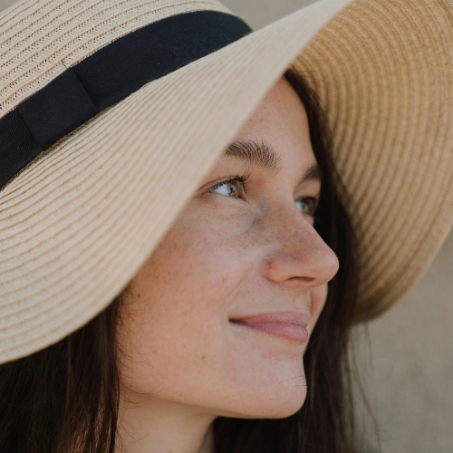
After keratosis has been treated, the skin remains more vulnerable. Prevention, therefore, does not mean giving up outdoor activities, but rather following a consistent protection plan. A wide-brimmed hat, tightly woven clothing, UV-protective fabrics, and sunscreen with high UVA and UVB protection help reduce exposure.
Avoid intense midday sun, tanning beds, and repeated sunburns. Check your forehead, nose, ears, lips, the backs of your hands, and scalp regularly in the mirror. Ask a trusted friend or family member to help you check your back or scalp. If you feel any rough patches of skin, make a mental note of the spot and monitor it over the next few weeks.
At ROC, prevention is part of a holistic approach: skin health, regeneration, and lifestyle go hand in hand. Those who have skin changes checked early often have access to gentler treatment options and avoid unnecessary anxiety.
The Regenerative Center in Aschheim is an interdisciplinary facility. In the field of dermatology and aesthetic medicine at ROC Derma, the focus is on skin diagnostics, treatment planning, and personalized care.
If actinic keratosis is suspected, we don’t just examine the specific area. We consider your overall risk profile: your history of UV exposure, skin type, medications, immune status, previous skin tumors, and which treatment fits best into your daily routine.
Would you like a medical evaluation of a rough, scaly, or recurring skin condition? Schedule an appointment at ROC Derma in Aschheim, near Munich. The sooner we can diagnose keratosis, the more targeted the treatment will be.