Bone Density Measurement (DXA Scan)
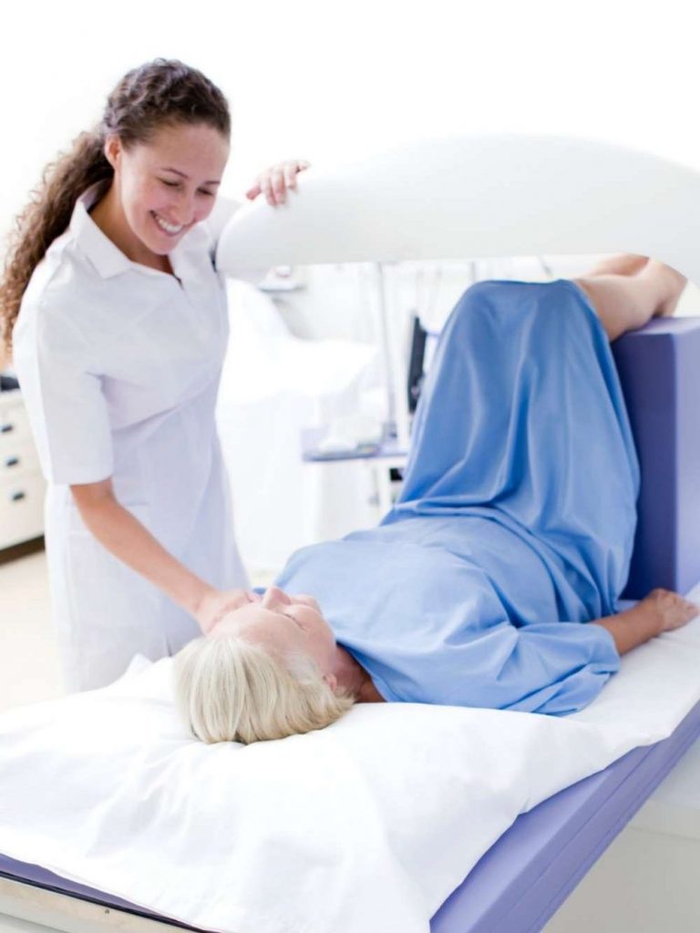
Bone density measurement using DXA (dual-energy X-ray absorptiometry), also known as osteodensitometry, is the recommended standard procedure for assessing bone density. It is used in particular when there is an increased risk of osteoporosis or osteoporosis-related bone fractures. At the ROC in Aschheim near Munich, we offer this precise examination to detect bone loss at an early stage and initiate targeted preventive or treatment measures. The measurement results are not considered in isolation but are medically evaluated in conjunction with potential risk factors, pre-existing conditions, medications, and previous bone fractures.
What is osteoporosis?
Osteoporosis is a chronic disease of the skeleton in which bone density and structure gradually decrease. As a result, the bones become porous, unstable and more susceptible to fractures - particularly in the hip, spine and wrist.
The condition often develops over a long period of time without any obvious symptoms. Possible signs may include a bone fracture following a relatively minor impact, a loss of height, an increasingly hunched back, or sudden onset of back pain.
Causes and risk factors for osteoporosis
Osteoporosis occurs when the natural balance between bone formation and bone resorption is disturbed. From around the age of 35, bone resorption slowly outweighs bone formation. In osteoporosis, however, this process is accelerated.
Common risk factors are
- Genetics: A family history increases the risk.
- Age & gender: Women after the menopause are particularly at risk as oestrogen levels fall.
- Lifestyle: Lack of exercise, insufficient calcium and vitamin D intake as well as smoking and excessive alcohol consumption promote bone loss.
- Diseases & medication: Rheumatism, hyperthyroidism, diabetes or long-term cortisone therapy can promote osteoporosis.
Symptoms - osteoporosis often goes unnoticed
Osteoporosis develops gradually and often remains undetected for a long time because it initially causes no pain. Only when the bones are severely weakened can it lead to:
- Back pain due to vertebral body fractures,
- changes in posture (hunchback, loss of height),
- Spontaneous bone fractures without major stress.
Early diagnosis is therefore crucial!
When is a bone density measurement useful?
A DXA scan may be advisable if there is an increased risk of osteoporosis or fracture. Possible reasons or risk factors include:
- a broken bone following a relatively minor fall
- known osteoporosis or osteopenia
- advanced age
- A family history of osteoporosis or hip fractures
- long-term treatment with cortisone
- inflammatory rheumatic diseases
- certain hormonal or metabolic disorders
- Being underweight or malnourished
- a confirmed vitamin D deficiency, especially when combined with other risk factors
- Smoking or heavy alcohol consumption
- repeated falls
- a significant decrease in height
- Hunchback or Suspected Vertebral Fractures
- Monitoring of previously treated osteoporosis
Whether a bone density test is medically appropriate in an individual case is determined based on the person’s overall risk profile.
How does the bone density measurement work?
Bone density testing (DXA scan) is painless and usually takes only a few minutes. During the test, you will lie relaxed on an examination table. The DXA machine uses low-dose X-rays to measure bone density in the lumbar spine and hip.
The examination consists of four steps:
- First, we'll discuss potential risk factors, pre-existing conditions, medications, and previous bone fractures.
- You will then be positioned appropriately on the examination table.
- The DXA machine measures bone density in the designated areas of the body.
- The measurements are then evaluated by a doctor and assessed in relation to your individual fracture risk.
What do T-score and Z-score mean?
The results of bone density measurements are expressed, among other things, as T-scores and Z-scores.
The T-score compares the measured bone density to the average bone density of young, healthy adults. It is used in particular to assess bone density in postmenopausal women and in men aged 50 and older.
For simplicity, the values are classified as follows:
- T-score up to −1.0: Bone density within the normal range
- T-score between −1.0 and −2.5: reduced bone density or osteopenia
- T-score of −2.5 or lower: a diagnostic indicator of osteoporosis
The Z-score compares bone density to the values of people of the same age and gender. It is particularly important for younger patients.
However, a single measurement alone does not determine whether treatment is necessary. The doctor’s assessment also takes into account age, previous bone fractures, comorbidities, medications, falls, and other risk factors.
Osteopenia or Osteoporosis: What's the Difference?
Osteopenia refers to a bone density that is below the normal range but has not yet reached the diagnostic threshold for osteoporosis. It does not automatically require treatment.
The overall risk of fracture is the key factor. Two people with the same T-score may be assessed differently based on their age, preexisting medical conditions, or previous bone fractures.
In cases of osteoporosis, bone density is significantly reduced. Here, too, the next steps are determined not solely by the T-score, but by the individual’s medical situation.
Why is early diagnosis of osteoporosis important?
As osteoporosis remains symptom-free for a long time, early diagnosis is crucial. It enables:
- Prevention of fractures through early treatment
- Individual therapy planning with diet, exercise and medication recommendations
- Monitoring bone health for an active life
If osteoporosis is not recognized and treated early, serious bone fractures can occur, even during everyday movements or minor falls. Femoral neck fractures are particularly common as a result of osteoporosis and can lead to serious physical and psychological consequences. Every year, around 160,000 people in Germany suffer a femoral neck fracture - a number that is expected to increase in the coming years due to rising life expectancy. A femoral neck fracture often leads to chronic pain, restricted mobility and loss of independence. Many patients are dependent on outside help and can no longer actively participate in life.
To prevent such serious consequences, bone density measurements should be carried out regularly in at-risk groups.
Supplementary Diagnostic Tests for Osteoporosis
A DXA scan measures bone mineral density but does not identify all possible causes of reduced bone strength. Depending on the results, further testing may therefore be advisable.
These include, for example:
- a comprehensive assessment of risk factors
- the physical examination
- the evaluation of previous X-ray, CT, or MRI images
- Imaging Tests for Suspected Vertebral Fractures
- the assessment of muscle strength, balance, and fall risk
- Laboratory Tests of Bone Metabolism
Depending on the clinical question, our orthopedic laboratory diagnostics can measure vitamin D, parathyroid hormone, and other relevant laboratory parameters, among other things.
What happens after the bone density test?
After the test, you will receive a medical assessment of your results. We will discuss whether your bone density is within the normal range, whether you have osteopenia, or whether there are signs of osteoporosis.
Depending on your personal risk of fracture, different next steps may be appropriate. These include, for example:
- an adequate intake of calcium and vitamin D
- Customized strength and balance training
- Measures to Prevent Falls
- the treatment of any underlying medical conditions
- drug therapy for osteoporosis when indicated
- a follow-up visit
For more information on the available treatment options, please see our guide to bone density testing and osteoporosis treatment.
Bone Density Testing at ROC Ortho
At the ROC, we combine DXA measurements with an orthopedic assessment of an individual’s fracture risk. If necessary, additional laboratory tests, imaging studies, or functional tests can be directly incorporated into the diagnostic process.
This way, you'll receive not only individual test results, but also a medical assessment and a recommendation for next steps.
Arrange your appointment for bone density measurement at the ROC in Aschheim near Munich!
E-Mail: praxis@roc-aschheim.de
Tel: +49 89 46259930
Our osteoporosis expert,
Martina Berger, looks forward to hearing from you!
