Digital volume tomogram (DVT)
High-resolution 3D X-ray diagnostics
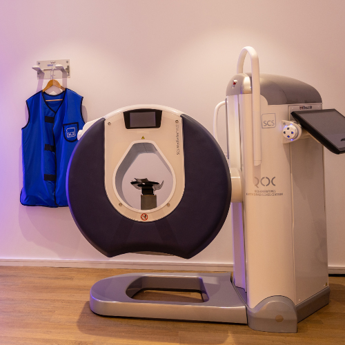
Digital volume tomography (DVT) allows for three-dimensional, high-resolution imaging of bones and joints. At ROC Ortho, this technique enables us to perform a precise assessment of bone structures and can serve as a valuable complement to conventional X-rays, particularly in complex cases.
A particular advantage in orthopedic diagnostics: Depending on the body region being examined, images can be taken under natural loading conditions—for example, while standing. This allows us to assess specific joint positions and bone changes as they appear under the stresses of everyday life.
At ROC Ortho in Aschheim, near Munich, we have access to DVT right here in our office. This allows us to closely integrate imaging, discussion of findings, and further treatment planning.
What is a digital volume tomogram (DVT)?
Digital volume tomography is a three-dimensional X-ray technique. While conventional X-rays produce two-dimensional images, DVT generates a three-dimensional dataset of the examined region. From this dataset, cross-sectional images in various planes as well as three-dimensional representations can be reconstructed. This allows us to view bony structures from different perspectives without having to take a new image for each view.
In orthopedics, DVT imaging is particularly well-suited for evaluating conditions affecting the extremities and joints that require a detailed visualization of bony structures.
When is a DVT useful in orthopedics?
Three-dimensional imaging is not necessary for every orthopedic condition. We determine whether a DVT is appropriate based on your symptoms, a physical examination, and, if applicable, any existing test results.
A DVT test may be considered, for example, in the following cases:
- unclear bony changes,
- complex joint configurations,
- Suspected specific fractures or minor bone injuries,
- Symptoms following injuries,
- degenerative changes and osteoarthritis,
- Malpositions and axial deviations,
- the assessment of joint positions under load,
- selected issues before proceeding further.
There is no one-size-fits-all answer to which imaging modality is best suited for your symptoms. Depending on the specific issue, digital X-rays, CT scans, ultrasound, or MRI can provide different types of information.
Which areas of the body can be examined using DVT?
DVT is particularly well-suited for high-resolution imaging of bony structures in various joints and extremities. Depending on the clinical question, areas of the arms and legs, among others, can be examined. The procedure is particularly useful when small bony structures, joint surfaces, or the position of a joint need to be assessed in detail. Before each examination, we determine whether DVT is, in fact, the appropriate imaging modality for the specific body region and clinical question.
DVT During Exercise: Examining Joints While Standing
A distinctive feature of digital volume tomography, when applied to suitable areas of examination, is the ability to take images under load. This can be relevant, for example, when evaluating conditions affecting the foot, ankle, or other weight-bearing joints. This is because certain misalignments or changes in joint position appear differently under the patient’s own body weight than when the patient is lying down. An image taken while standing can therefore provide us with additional information about how bone structures and joints behave under realistic loading conditions. This information can be particularly helpful for further diagnosis and treatment planning, especially for symptoms that occur mainly while standing or walking.
DVT or X-ray: What's the difference?
Conventional X-rays are among the most important imaging techniques in orthopedics. They provide two-dimensional images and are suitable for diagnosing a wide range of conditions affecting bones and joints.
In contrast, DVT generates a three-dimensional data set. This allows bone structures to be visualized without overlap and in various cross-sectional planes.
Which imaging test is more appropriate depends on the specific issue. For many conditions, a conventional X-ray is perfectly sufficient. However, if we need to evaluate a complex bony structure in greater detail or require a three-dimensional view, a CT scan can provide additional information. Our goal, therefore, is not to use a CT scan as often as possible, but to select the imaging modality that is best suited to your condition.
DVT or CT: What's the difference?
Both digital volume tomography and computed tomography (CT) provide three-dimensional and cross-sectional images, respectively.
DVT specializes in specific areas of examination and, in particular, in high-resolution imaging of bony structures. The examination can be performed quickly and, depending on the area, also allows for images to be taken under stress.
A CT scan, on the other hand, has a much broader range of diagnostic applications and, depending on the clinical question, can also examine soft tissues and larger areas of the body.
Similarly, with regard to radiation exposure, it is not possible to specify a general percentage value for every DVT and CT scan. The actual radiation dose depends, among other things, on the area being examined, the equipment used, and the specific imaging protocol. We therefore assess on a case-by-case basis which procedure is appropriate and necessary for the medical issue at hand.
DVT or MRI: When Should Which Test Be Used?
DVT and MRI address different diagnostic questions.
DVT is particularly well-suited for detailed imaging of bony structures. MRI, on the other hand, primarily provides information about soft tissues such as cartilage, ligaments, tendons, muscles, and other structures. Therefore, depending on the specific clinical question, either one or the other imaging modality may be appropriate for a joint problem. In certain cases, the two imaging modalities complement each other.
At ROC Ortho, we can combine various diagnostic procedures. In addition to DVT, we have access to digital X-rays, high-resolution ultrasound, and open MRI, among other options. This allows us to select the appropriate imaging modality based on the specific structure we wish to evaluate.
Advantages of Digital Volume Tomography
When used to address appropriate clinical questions, DVT offers various advantages for orthopedic diagnosis:
- three-dimensional representation of bony structures,
- high-resolution cross-sectional images,
- Examining the region under study from various perspectives,
- short recording time,
- Depending on the body region, an examination under natural stress may be possible,
- immediate availability at the ROC,
- Direct integration of the images into orthopedic diagnosis and treatment planning.
However, the medical indication is always the deciding factor. Not every symptom requires three-dimensional imaging.
How is a DVT test performed at the ROC?
Before the examination, we will discuss your symptoms and determine which diagnostic questions need to be addressed. Any existing X-ray, CT, or MRI images can provide important information. For the actual DVT scan, the area of the body to be examined will be positioned accordingly. Depending on the specific diagnostic question, the examination may also be performed under stress. The scan itself takes only a short time. During the scan, you should keep the affected area as still as possible to ensure good image quality. Afterward, the cross-sectional images and 3D renderings can be evaluated. Since CT is available directly at ROC Ortho, we can incorporate the results into further orthopedic diagnostics and treatment planning.
DVT Diagnostics at ROC Ortho in Aschheim near Munich
For us, high-quality imaging is not an end in itself. What matters most is that the examination answers a specific medical question. At ROC Ortho, we therefore combine a physical examination with the most appropriate imaging modality for each case. Digital volume tomography (DVT) complements our diagnostic capabilities, particularly when we need to assess bony structures in three dimensions and in detail. Since, in addition to DVT, we have access to other techniques such as digital X-rays, ultrasound, and open MRI, we can determine—based on your symptoms—which method will provide the necessary information. On this basis, we will discuss the findings with you in a clear and understandable manner and plan the next diagnostic or therapeutic steps.
ROC Ortho
Private Orthopedic Practice
St.-Emmeram-Str. 5
85609 Aschheim
Email: praxis@roc-ortho.de
Phone: +49 89 46259930
Opening hours
| Monday | 7:30 a.m. – 6:00 p.m. |
| Tuesday | 7:30 a.m. – 6:00 p.m. |
| Wednesday | 7:30 a.m. – 8:00 p.m. |
| Thursday | 7:30 a.m. – 6:00 p.m. |
| Friday | 7:30 a.m. – 3:00 p.m. |
Frequently Asked Questions About Computed Tomography (CT) Scans
What is a DVT?
DVT stands for digital volume tomography. It is a three-dimensional X-ray imaging technique that can be used to produce high-resolution images of bone structures in particular.
What can you see on a DVT scan?
A CT scan provides detailed images of bones, joint surfaces, and other bony structures. The three-dimensional data set allows us to view the examined region from different angles.
What is the difference between a DVT and a regular X-ray?
A conventional X-ray produces two-dimensional images. CT scans, on the other hand, generate a three-dimensional data set from which various cross-sectional images and views can be reconstructed. For many diagnostic purposes, a standard X-ray is sufficient; however, in cases of more complex bony abnormalities, a CT scan can provide additional information.
What is the difference between a DVT and a CT scan?
Both procedures use X-rays and provide cross-sectional images or three-dimensional representations. DVT is specifically designed to provide high-resolution images of certain bony structures. A CT scan has a broader range of diagnostic applications. Which procedure is appropriate depends on the specific medical issue at hand.
How high is the radiation exposure associated with DVT?
The radiation exposure from a DVT scan cannot be expressed as a universal value. It depends, among other things, on the body region being examined, the equipment, and the imaging protocol used. For this reason, a DVT scan should only be performed when medically indicated.
How long does a DVT test take?
The actual scan takes only a short time. In addition, there is the preparation, the proper positioning of the body part being examined, and the subsequent evaluation of the images.
Can a DVT be performed while standing?
Depending on the body region being examined and the clinical question, DVT imaging can be performed under load. This allows for the assessment of specific joint positions and bony relationships as they appear under the patient’s own body weight.
Is it possible to have a DVT even if you have a fear of enclosed spaces?
The examination does not take place in a narrow tube, as is the case with a traditional MRI. For this reason, the open scanning environment can be particularly comfortable for patients who feel uncomfortable during examinations in confined spaces.